About The Procedure
Radioembolization can be used in combination with chemotherapy for more effective treatment. The source of the radiation, Yttrium-90 (Y90) is baked into tiny resin beads that delivered to the tumors through a catheter placed in the hepatic artery, which is the source of blood flow to the tumors. The radioactive beads remain in the tumors, blocking blood flow and shrinking it without exposing the rest of the body to the radiation.
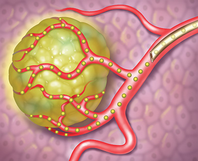
Side effects are generally minimal because of the positioning of the beads, and patients go home within hours after the procedures. Over a period of about a month, the radiation in the beads is depleted and any remaining particles can safely remain in place.
Radioembolization is primarily used to treat cancers of the liver that have metastasized or spread from other locations in the body, especially the colon. It can be used in combination with chemotherapy or surgery, and can be used to shrink large tumors to allow for surgical removal. This therapy is not appropriate in patients with severe liver disease or in patients who have abnormal blood flow between the liver and the lungs.
What to Expect
If your doctor decides that you are a candidate for this procedure, it is performed in a hospital by CRA Interventional Radiologists. The procedure is performed on two separate procedure days. Each procedure day, medical staff will insert an IV and connect you to monitors to measure your vital signs and you’ll receive medication to keep you comfortable during the procedure. The radiologist will insert a catheter into the groin artery and guide it into place in your liver. You may feel some pressure during the procedure, but no pain.
The mapping procedure is scheduled several days prior to the actual embolization or treatment procedure. The Interventional Radiologist will map your liver anatomy to identify arteries that may be feeding the tumors. They also look for small arteries near the tumors that may lead to the stomach or intestines and-if necessary- block these arteries to keep radiation from reaching these organs. Macro aggregated albumin (MAA) particles are injected into your liver to trace where the particles go, and a scan is performed to make sure they stay in the liver and the tumors being treated.
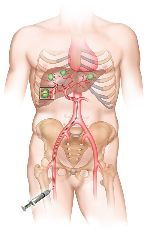
In the second procedure, the catheter is again placed directly into the liver artery and the radioactive beads are delivered. A follow-up scan will be performed to confirm the beads were directly delivered into the tumors. Each of the procedures takes 60-120 minutes.
After each treatment, the catheter in your groin is removed and you’ll be asked to lie flat for a few hours. You may experience some nausea, pain, or fever after the procedure and for the next few days. Medications will be prescribed to manage any side effects.
After you are sent home from the procedure, you should avoid driving for 24 hours, limit activities, and avoid heavy lifting or physical exertion for 2-3 days. Full recovery usually takes 7-10 days. You will be seen for follow-up one week after the procedure. If you have tumors in both lobes of your liver, you may need a second round of treatment scheduled a month apart.
Contact us for more information: 315-234-4686.